Whether a Malamute Gets Corneal Dystrophy
It's truly unsettling to notice a cloudy film or an unusual opacity developing in your beloved Alaskan Malamute's eye. Immediately, your mind jumps to all sorts of scary possibilities, and the thought of their vision being compromised is heartbreaking. You might wonder if it's an injury, an infection, or something much more serious. This comprehensive guide is here to shed light on one specific condition that can cause this hazy appearance: corneal dystrophy in Alaskan Malamutes, offering clarity on what it is, how it's diagnosed, and what it means for your furry family member.
Understanding Corneal Dystrophy in Alaskan Malamutes
When you look into your Malamute's intelligent, often dark brown eyes, you expect to see a clear window to their soul. But sometimes, a subtle change starts to appear, a cloudiness or a crystalline sparkle that wasn't there before. This can be the first sign of corneal dystrophy, a condition that, while it sounds alarming, isn't usually as dire as other eye issues. It's a genetic puzzle piece in the Malamute world, one that responsible owners and breeders strive to understand.
What Exactly Is Corneal Dystrophy?
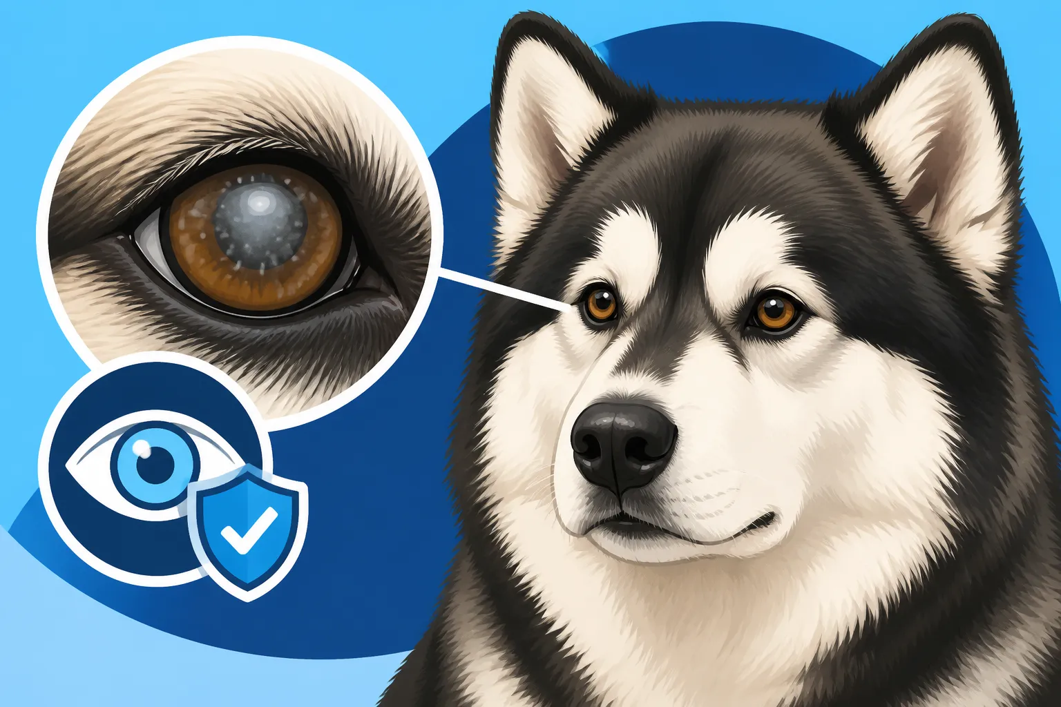
Corneal dystrophy is a non-inflammatory, often inherited eye condition characterized by the deposition of abnormal material within one or more layers of the cornea. Think of the cornea as the transparent outer layer at the very front of your dog's eye, much like the crystal on a watch. Its job is to protect the eye and to help focus light onto the retina, which is why its clarity is absolutely paramount for good vision. When these deposits form, they can make the cornea appear hazy, opaque, or even sparkling with tiny crystals.
There are generally three types of corneal dystrophy, named for the layer of the cornea they affect: epithelial (outermost layer), stromal (middle, thickest layer), and endothelial (innermost layer). For Alaskan Malamutes, the stromal type is most commonly observed. This form typically involves the accumulation of lipids (fats) or cholesterol crystals within the corneal stroma, creating those characteristic grayish-white or crystalline opacities. Unlike inflammatory conditions, corneal dystrophy doesn't usually cause redness, swelling, or pain in the eye, at least not in its primary form. It’s more about the structural change to the cornea itself.
It’s crucial to understand that while it's progressive, corneal dystrophy in Malamutes often advances very slowly and rarely leads to blindness or significant discomfort. This sets it apart from more acute or painful conditions like corneal ulcers, which demand immediate veterinary attention. The deposits are typically bilateral, meaning they affect both eyes, though they might not appear simultaneously or be perfectly symmetrical in their presentation or severity.
How Corneal Dystrophy Manifests in Malamutes
The tell-tale sign of corneal dystrophy in an Alaskan Malamute is the appearance of a cloudy, hazy, grayish-white, or even shimmering crystalline patch within the cornea. These opacities can vary in size and shape, sometimes appearing as discrete dots, rings, or diffuse areas. You might first notice them when your Malamute is in a certain light, or perhaps during a routine cuddle session where you're looking closely at their eyes. These changes are usually not accompanied by squinting, tearing, or any signs of pain, which can be a relief but also makes early detection tricky if you're not looking carefully.
Often, these deposits will be found in the central part of the cornea, but they can also appear in the periphery. For many Malamutes, the condition presents as an oval or circular area of white-to-gray opacity in the center of the cornea. It’s important to note that while the condition is considered hereditary, the exact age of onset can vary widely among individuals, from early adulthood to middle age. The deposits also tend to be quite stable; they might slowly enlarge over years, but rapid changes are uncommon.
One of the most reassuring aspects for Malamute owners is that these deposits, even when quite noticeable, generally do not impede vision to a significant degree for the dog’s normal activities. Because Malamutes are incredibly adaptable and often rely on their keen sense of smell and hearing, minor visual impairments might go unnoticed by owners for a long time. However, any change in your dog’s eye, no matter how subtle, warrants a visit to your veterinarian to rule out other, potentially more serious, issues.
Distinguishing Corneal Dystrophy from Other Eye Conditions
Spotting a cloudy eye in your Malamute is always a cause for concern, and rightly so. But it's important not to jump to conclusions, as many conditions can present with similar visual cues. Understanding the nuances between corneal dystrophy and other eye ailments is essential for seeking the correct diagnosis and ensuring your Malamute receives appropriate care.
Cloudy Eyes: When It's Not Corneal Dystrophy
While corneal dystrophy causes a cloudy appearance, it's vital to recognize that other eye conditions can mimic this symptom, some of which are much more urgent or severe. For instance, cataracts are a very common cause of cloudiness, but they affect the lens of the eye, not the cornea. The lens is located behind the iris (the colored part of the eye), so a cataract will typically appear as an opaque spot deeper within the eye, rather than on the very surface. For more on this, you might explore content on Cataracts in an Alaskan Malamute.
Another common age-related change is nuclear sclerosis, which presents as a bluish-gray haze in the center of the lens in older dogs. This is a normal aging process where the lens fibers harden and compress; it doesn't significantly impair vision and is distinct from cataracts. Then there are inflammatory conditions like keratitis (inflammation of the cornea) or corneal ulcers, which cause cloudiness but are usually accompanied by redness, pain (manifesting as squinting or pawing at the eye), discharge, and a sudden onset. These require immediate veterinary intervention to prevent permanent damage or vision loss. Glaucoma, characterized by increased pressure within the eye, can also lead to a cloudy cornea, but it’s often very painful and causes the eye to appear enlarged or bloodshot. Given the range of possibilities, a professional diagnosis is non-negotiable.
The Importance of a Proper Diagnosis
When you notice any changes in your Malamute's eyes, the first and most critical step is to schedule an appointment with your veterinarian or, ideally, a veterinary ophthalmologist. They possess the specialized tools and expertise to accurately diagnose the condition. The diagnostic process typically begins with a thorough ophthalmic examination, which involves using specialized lights and magnifiers to carefully inspect all parts of the eye. Your vet will look for the precise location, size, and character of the opacities.
A slit lamp biomicroscopy is a common tool used by ophthalmologists, allowing for a highly magnified, three-dimensional view of the cornea's layers to pinpoint exactly where the deposits are located. Other tests, such as fluorescein staining, might be performed to rule out corneal ulcers, as the dye will stick to any areas where the corneal surface is damaged. In rare cases, if the diagnosis is ambiguous, corneal scrapings or biopsies might be taken for histopathological examination, but this is uncommon for typical corneal dystrophy. Early and accurate diagnosis not only ensures your Malamute gets the right care but also gives you peace of mind, especially when ruling out more aggressive or painful conditions. This thorough evaluation is why having pet insurance, like a plan from Healthy Paws, can be incredibly valuable, as it helps cover the costs of these specialized diagnostic tests.
Managing and Living with Corneal Dystrophy
Discovering your Alaskan Malamute has corneal dystrophy can be a worry, but understanding the usual prognosis and management strategies can alleviate a lot of that stress. For most Malamutes, this is a condition that requires observation rather than intensive treatment, allowing them to lead full, happy lives.
Treatment Options and Prognosis
Here’s the good news for the majority of Alaskan Malamute owners: most cases of corneal dystrophy do not require active treatment. The condition is largely asymptomatic, meaning it doesn't cause pain, discomfort, or significant vision impairment for the dog. The lipid or cholesterol deposits simply sit there, usually progressing so slowly over many years that your Malamute adapts without issue. Regular monitoring by your veterinarian is typically all that's needed to track any changes.
However, in very rare instances, the deposits might become extensive enough to truly impact vision, or they could lead to secondary complications like recurrent corneal erosions (superficial ulcers). If this happens, a veterinary ophthalmologist might consider options such as topical lubricating eye drops to protect the corneal surface, or hypertonic saline drops to try and reduce corneal swelling. Surgical intervention, like a superficial keratectomy (where the superficial affected layers of the cornea are carefully removed), is exceedingly rare and generally reserved for cases where vision is severely compromised or painful ulcers are recurrent. The prognosis for most Malamutes with corneal dystrophy is excellent for a normal quality of life, with minimal intervention required. Even if surgery were ever deemed necessary, a comprehensive pet insurance plan from Healthy Paws could help manage the financial burden.
Genetic Considerations and Breeding
Corneal dystrophy in Alaskan Malamutes is widely considered an inherited condition. This means it's passed down through generations, making genetic screening and responsible breeding practices incredibly important. While specific DNA tests for Malamute corneal dystrophy are not yet widely available or standardized across all lines, reputable breeders are diligent about monitoring their breeding stock for any signs of the condition and will generally remove affected dogs from their breeding programs. They understand the importance of genetic health and are often proactive about all The Health Tests Both Alaskan Malamute Parents Should Have.
If your Malamute is diagnosed with corneal dystrophy, it is strongly advised that they not be used for breeding. Even if the condition is mild in an individual, they carry the genes that could pass it on to future generations, potentially leading to more severely affected offspring or increasing the prevalence in the breed. Similarly, breeding carriers (dogs that carry the gene but don't show symptoms) should be approached with extreme caution, ideally only when bred to clear individuals, as detailed in discussions like Whether a Carrier Malamute Can Be Bred. This commitment to genetic health helps ensure the future vitality of the breed and minimizes the suffering caused by preventable inherited diseases.
Supporting Your Malamute's Eye Health
Beyond addressing specific conditions like corneal dystrophy, maintaining overall eye health for your Alaskan Malamute is an ongoing part of responsible pet ownership. Proactive observation and a supportive lifestyle can make a significant difference in their well-being throughout their life.
Routine Care and Observation
One of the most powerful tools you have as a Malamute owner is diligent, routine observation of your dog's eyes. Make it a habit to glance at them daily, perhaps during playtime or grooming. Look for clarity, brightness, and any signs of discomfort. What you're watching for includes: changes in the normal transparency of the eye (like new cloudiness or opacities), any redness in the white parts of the eye, unusual discharge (clear tearing is sometimes normal, but thick or colored discharge is not), persistent squinting, frequent rubbing or pawing at the eyes, or any behavioral changes that might suggest vision problems, such as bumping into furniture or being hesitant to navigate familiar spaces.
Should you notice any of these symptoms, especially if they appear suddenly, it's critical to contact your veterinarian without delay. Even if it turns out to be something minor, prompt veterinary attention can prevent a small issue from becoming a major one, particularly when dealing with the delicate structures of the eye. Regular veterinary check-ups are also crucial, as your vet can catch subtle changes you might miss. Many veterinarians recommend a yearly eye exam, especially as your Malamute ages, to monitor for conditions like nuclear sclerosis or progressive retinal atrophy (Whether a Malamute Gets Progressive Retinal Atrophy).
The Role of Nutrition and Supplements
When it comes to corneal dystrophy specifically, it’s important to understand that it's primarily a genetic condition, not one caused by nutritional deficiencies or excesses in typical circumstances. Therefore, no special diet or supplement is known to cure or prevent it. However, a well-balanced, high-quality diet is always fundamental for overall health, including eye health. Ensuring your Malamute receives adequate vitamins and minerals supports all bodily functions, including maintaining healthy ocular tissues.
Some general eye health supplements for dogs contain antioxidants like Vitamin C, Vitamin E, lutein, and zeaxanthin, which are believed to help protect cells from oxidative stress. While these might support overall eye wellness, it's important to reiterate they are not a "treatment" for corneal dystrophy itself. Always consult your veterinarian before adding any supplements to your Malamute's diet, as excessive amounts of certain nutrients can be harmful. For example, while zinc is vital, too much can be toxic, and some Malamutes have specific Whether a Malamute's Zinc Problem Is Diet or Genetics issues. Focusing on a complete and balanced diet, tailored to your Malamute's age, activity level, and any other health conditions, is the best approach. Avoid feeding your Malamute any Foods That Are Dangerous to an Alaskan Malamute which could lead to systemic problems that indirectly impact eye health.
| Eye Condition | Primary Affected Part | Appearance (Typical) | Pain Level (Typical) | Urgency of Vet Visit (Typical) |
|---|---|---|---|---|
| Corneal Dystrophy | Cornea (surface) | Grayish-white, crystalline opacity | None | Routine/Monitoring |
| Cataract | Lens (inside eye) | Opaque spot deep within pupil | None | Routine/Monitoring |
| Nuclear Sclerosis | Lens (inside eye) | Bluish-gray haze, uniform | None | Low (Normal Aging) |
| Corneal Ulcer | Cornea (surface) | Hazy spot with redness, indentation | High | Emergency |
| Keratitis | Cornea (surface) | Hazy, often with redness, discharge | Moderate | Urgent |
| Glaucoma | Entire eye | Enlarged, cloudy, bloodshot eye | High | Emergency |
Disclaimer
The information provided in this article is for general informational purposes only and does not constitute veterinary advice. Always consult with a qualified veterinarian or veterinary ophthalmologist for any health concerns or before making any decisions related to your Alaskan Malamute's health and treatment.